Case Four – April 2017
- 76 year old female presented with a scaly patch on the left areola.
- No known history of breast cancer.
- The lesion was unresponsive to topical corticosteroids.
- Contact dermatitis
- Paget disease

There is an intraepidermal glandular proliferation composed of well-formed tubules present throughout the epidermis.
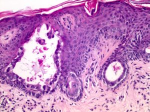
The tubules are lined by atypical epithelial cells with vacuolated cytoplasm, hyperchromatic nuclei, and inconspicuous nucleoli. Scattered mitotic figures are present.
Immunohistochemistry:
- The intraepidermal glands are strongly positive for Cam5.2
Glandular Mammary Paget Disease
- This is an extremely rare variant of mammary Paget disease in which the tumor cells form well-defined glands within the epidermis.
- This variant may also be called acinar mammary Paget disease.
- This variant is unique since it is composed predominantly of intraepidermal glands. In contrast, conventional mammary Paget disease typically shows discohesive, single atypical cells with foamy, vacuolated cytoplasm in a pagetoid array throughout the epidermis.
- The tumor cells in the glandular variant of mammary Paget disease are positive for the same immunohistochemical stains as conventional mammary Paget disease, including CEA, EMA, CK7, Cam5.2 and cytokeratin AE1/AE3.